
The Ultimate History of CAD/CAM in Orthotics
, by Hugh Sheridan, 8 min reading time
, by Hugh Sheridan, 8 min reading time
The prevalence of CAD/CAM technologies in P&O has grown over the past decades with the advent of new scanners, modification softwares, 3D carvers, 3D printers, and printing materials.
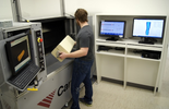
CAD/CAM was introduced to P&O over three decades ago. One of the first reports of CAD/CAM, published in 1985, described a “software package” for the manufacture of transtibial sockets[2]. The second author of this publication went on to develop Vorum, the first and longest-standing CAD/CAM company dedicated to P&O. Vorum was initially focused on 3D carvers, which uses a milling machine to carve a foam block based on a CAD drawing[3].
The first attempts of 3D printing in a P&O-specific application were reported in the early 1990s, about a decade after the first published patent of any 3D printing technology. These studies described the fabrication of transtibial socket using stereolithography (SLA), and fused deposition modelling (FDM) with the Squirt-Shape™ printer.[4]
The prevalence of CAD/CAM technologies in P&O has grown over the past decades with the advent of new scanners, modification softwares, 3D carvers, 3D printers, and printing materials. Though many view 3D scanning and printing as a way to reduce costs and increase access to P&O devices, CAD/CAM is not always synonymous with lower cost.[5]
Current literature indicates a steady increase in adoption of CAD/CAM technologies, but not to the point of overtaking traditional methods. A 2021 study indicates increased interest in CAD/CAM from both developed and developing countries. However, developing countries have faced challenges in adoption such as accessibility, resources, qualified practitioners, and gaps in knowledge.[6] In the United States, the 2022 Practice Analysis indicates that 30% of prostheses incorporate CAD/CAM, increasing from 23% in the 2015 study[7]. Relative to prostheses, orthoses made with CAD/CAM are entering the field more slowly. However, they show promise as far as comfort, biomechanical benefits, and optimised material properties.[8]
Currently, CAD/CAM technologies are used in the fabrication of all kinds of diagnostic and definitive devices, including:
The process for creating a prosthesis or orthosis with CAD/CAM can be broken into three parts:[9]
For a practitioner to incorporate CAD/CAM into their practice, they do not have to use all three parts of the process. For example, a prosthetist may fit a 3D printed diagnostic socket made from a scan of a modified plaster model. An orthotist or technician may pull plastic (using traditional methods) over a carved foam model for a scoliosis brace.
Several types of scanners can be used to capture an impression of a patient’s limb or anatomical feature. Two commonly used technologies in P&O are:
Some are used independently as a handheld or stationary scanner, and some work in conjunction with an iPhone, iPad, or other device.
Several studies have confirmed the reliability of capturing limb shape compared to traditional methods.[11] [12][13][14]
In order for a scan to be turned into a printable prosthesis or orthosis, it must be modified using CAD software. While there are P&O-specific programs for digital modifications, some use other design programs such as Autodesk Meshmixer, Autodesk Fusion 360, Dassault Systemes SolidWorks, etc.
There are several platforms for performing modifications, including:
Before sending a modified scan to print or carve, the file must be prepared for the specific manufacturing technology. For 3D printing, this process is usually called “slicing.”[16] This can sometimes be performed in the same CAD program for modifications, but generally the design file is saved as a .stl or .obj file and imported to a separate slicing software.
There are many different 3D printing technologies, but three are predominantly used in P&O:[17]
The selected printer, print properties and material determine ultimate properties of the prosthesis and orthosis.
A systematic review by Kim et al. reports that transtibial sockets made with 3D printing show promise as definitive sockets based on ISO standard testing methods.[18]
In a survey of 250 P&O practitioners internationally, 97% report that adopting CAD/CAM positively impacts patient care and outcomes. However, 98% report that CAD/CAM is not yet well understood by P&O clinicians.[22] It is difficult to identify the clinical impact of CAD/CAM with the current lack of standardization and established workflow.[23]
Studies indicate that a digital workflow has the potential to improve fabrication efficiency.[9] One advantage is the ability to save an impression or mold on a computer vs. store within a fabrication lab. This allows duplication of devices and fast comparison between scans (such as a patient’s residual limb one year post-amputation vs. three months post-amputation).
For clinicians hesitant to incorporate 3D printed lower limb prostheses, they often question the overall strength and durability of 3D printed parts for lower limb prostheses.[18][9]According to a systematic review, it is currently impossible to directly compare the strength of a 3D printed socket to a traditional carbon laminated socket with existing testing methods. However, the strength of 3D printed sockets shows promise for use in definitive prostheses. Recommended design strategies to increase strength include reinforcing the distal end and/or using composite filaments.[18]
One study reported that transtibial sockets manufactured with CAD/CAM resulted in better quality of life than those manufactured by traditional methods.[24]
Some groups are trying to use CAD and 3D printing to improve access to P&O worldwide. One group defined a method to provide 3D printed prostheses to patients in Sierra Leone in order to address an unmet global health need.[25] A different group cites 3D printing as a way to incorporate low cost materials and rapid prototyping to increase access to prostheses.[6]
Most of the research analysing CAD/CAM for upper limb (UL) prostheses has focused on paediatrics. 75% of the prostheses developed for children were made using FDM. A review of 3D printed UL prostheses reported that evidence regarding durability, functionality and user acceptance is lacking.[5]
One study found that ankle-foot-orthoses (AFOs) produced with AM are comparable to traditional AFOs in terms of spatio-temporal parameters. The review notes that only one of eleven studies conducted durability testing, and that sample sizes and study quality were generally low.[8]
3D scanning and AM have gained popularity particularly in the production of foot orthoses. A systematic review by Daryabor et al. reports that foot orthoses may improve comfort and biomechanics for those with flat feet, but indicates no statistical difference to those made with traditional methods.[26]
Custom 3D printed orthoses show promise for stabilising the upper limb for those with musculoskeletal conditions. Advantages in this area include improved aesthetics and the ability to create lightweight, well-ventilated orthoses. Current barriers to implementation are lack of training, skill, and high upfront costs for clinicians providing UL orthoses.[27]
CAD/CAM has been used for the fabrication of spinal orthoses for over two decades. The first central fabrication facility to incorporate CAD/CAM for scoliosis now has over 6,000 carved patient molds. They cite the following as advantages to scanning for scoliosis braces versus the traditional method of casting:[28]
3D printed scoliosis orthoses are relatively newer to the field than scanning, digital rectification, and carving. A 2022 randomized controlled trial compared the efficacy of 3D printed orthoses to conventional orthoses for the treatment of adolescent idiopathic scoliosis (AIS). Both groups had comparable in-orthosis correction and angle reduction after two years. This study also reports that the orthotist dedicated 4.8 hours less to the design and fabrication of the 3D printed orthoses. [29]